What Are X-rays?
In the world of modern medicine and diagnostics, X-rays have become one of the essential tools relied upon by doctors and healthcare professionals. But what are X-rays? How do they work? And what is their significance in diagnosing diseases and detecting health issues? In this article, we provide a comprehensive overview of X-rays, their mechanism of action, practical applications, and related safety considerations.
Definition of X-rays
X-rays are a type of electromagnetic radiation, similar in properties to visible light but with higher frequency and greater energy. Discovered in 1895 by German physicist Wilhelm Conrad Roentgen, who won the Nobel Prize in Physics in 1901 for his discovery, X-rays were first used to reveal broken bones, opening the door to extensive medical applications.
X-rays are naturally produced during processes in space, such as stellar explosions, but in medical applications, they are generated artificially using specialized devices. X-rays can be described as invisible radiation with the ability to penetrate materials and tissues, primarily used for imaging internal body structures and detecting changes not visible to the naked eye.
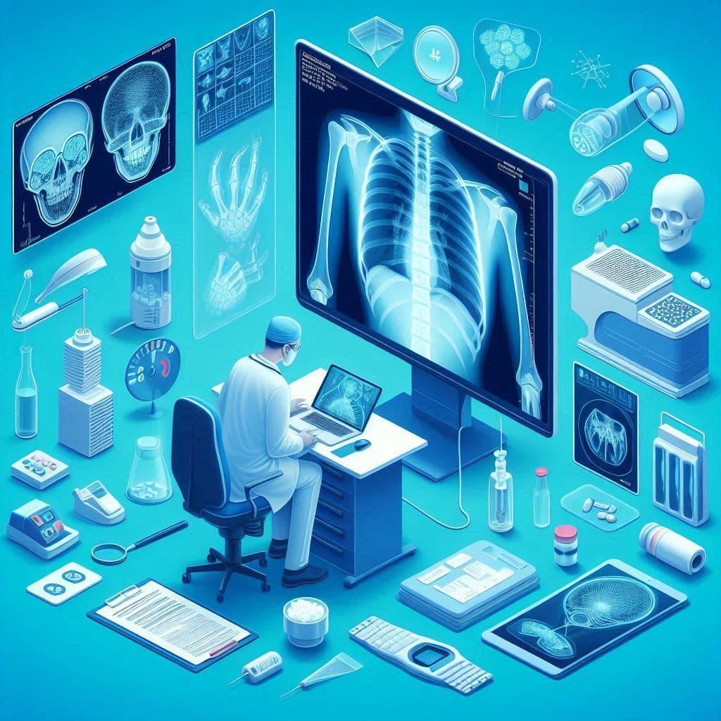
In the medical field, X-rays are used to obtain images of internal body tissues, aiding doctors in diagnosing diseases and injuries. This technology is widely used in medical imaging, including conventional X-rays, computed tomography (CT) scans, and radiographic imaging. X-rays help improve diagnostic accuracy and facilitate early patient treatment.
How X-rays Work
How X-rays Are Generated
X-rays are generated using a device called an X-ray tube. The tube consists of two main components: the cathode and the anode. The cathode is the source of electrons, while the tube contains a metallic anode that receives the electrons. When electrons collide with the anode, their energy is converted into X-rays.
The process begins with an electric current passing through the cathode, accelerating electrons towards the tube. When these electrons strike the anode, some of their energy is converted into X-rays. The X-rays are directed through the body to another receiving device, such as film or a digital detector, where the image is captured and recorded.
How X-rays Pass Through the Body
X-rays interact with tissues in the body differently depending on the density of the tissues. Dense tissues, like bones, absorb most of the X-rays, making them appear clearly on the image. In contrast, less dense tissues, such as soft tissues, allow more X-rays to pass through, making them appear darker on the image.
The ability of X-rays to penetrate tissues varies based on the characteristics of the tissues themselves. For instance, soft tissues like muscles and fat allow more X-rays to pass through compared to bones, resulting in them appearing darker in the final image. These contrasts in density help doctors analyze the images and identify any health issues that may be present.
Results and X-ray Imaging
The images produced by X-rays are used by doctors to diagnose various health problems, from fractures and injuries to chronic diseases like cancer. The images are displayed on a screen or printed on special films, where doctors can analyze them in detail to determine the patient’s condition and make appropriate treatment decisions.
In the case of fractures, X-rays clearly show the location and type of fracture, helping determine the best treatment. For diseases like infections or tumors, X-rays help identify the location and size of abnormal changes, making it easier for doctors to make correct treatment decisions.
Practical Applications of X-rays
In Medicine
In the medical field, X-rays are primarily used to diagnose various conditions. They help identify bone fractures, diagnose lung infections, and detect tumors. X-rays are valuable in emergency care, where they can quickly assess injuries and medical emergencies.
Additionally, X-rays are used in evaluating chronic conditions such as arthritis or heart disease. They also play an important role in monitoring treatment progress, allowing doctors to compare new images with previous ones to track changes in the patient’s condition.
In Industry
The use of X-rays is not limited to the medical field; it extends to industry as well. X-rays are used in material inspection and welding examination to detect defects and cracks. They are also employed in quality control to ensure the safety and suitability of products for use.
In heavy industries like aerospace and automotive manufacturing, X-rays are used to inspect materials and components for any defects that could impact performance or safety. They are also used in security to scan luggage at airports to identify contents without the need to open them.
In Research
X-rays play a significant role in scientific research, particularly in studying materials and tissues at the atomic level. X-rays are used in studying crystal structures and materials, contributing to advancements in science and technology.
In chemical and physical research, X-rays are employed to study material structures at the atomic level, aiding in the development of new materials and improving manufacturing processes. They are also used in medical research to understand how drugs affect tissues and cells.
Safety and Precautions
Potential Risks
Although X-rays provide significant medical benefits, excessive exposure can pose health risks. Overexposure to X-rays can increase the risk of cancer, making it important to use this technology carefully and according to health standards.
Potential risks also include short-term side effects such as redness or irritation of the skin in cases of overexposure. Additionally, it is emphasized that the risks and benefits should be evaluated for each case to minimize the impact of X-rays on patient health.
Safety Measures
Several safety measures are taken to reduce the risks associated with X-rays. These measures include using protective shields, limiting the number of X-rays required, and providing proper training for medical practitioners. Additionally, assessing the benefits versus risks is crucial before performing any X-ray examination.
Other safety procedures include ensuring that patients wear protective clothing if necessary and applying imaging techniques that minimize exposure to X-rays. Modern imaging technologies also rely on reducing the required dose to obtain accurate images.
Dokki Scan : A Leader in Providing X-ray Services
At Dokki Scan, we offer X-ray services with the highest standards of quality and precision. We use the latest technology and equipment to ensure accurate and reliable results. Our team of specialized doctors and technicians works diligently to provide excellent medical care and ensure patient safety.
We understand the importance of precise imaging in the diagnostic process, and we are committed to offering the best services in X-ray technology while maintaining the highest standards of safety and health precautions. Your visit to Dokki Scan guarantees you receive outstanding medical services based on advanced imaging techniques and extensive experience.
Additionally, we provide specialized consultations on how to prepare for examinations and ensure that patients are given clear and detailed information about procedures and expectations. We prioritize patient satisfaction and comfort, creating a welcoming and safe environment for everyone who comes to us.
Conclusion
X-rays are a vital tool in both medicine and industry, offering internal views of the body and materials, which helps in diagnosing diseases and detecting defects. By understanding how X-rays work and their applications, you can appreciate their role in healthcare and scientific research. At Dokki Scan , we take pride in delivering high-quality X-ray services with utmost safety, ensuring the best care for our patients.
For more information about our X-ray services or to schedule an appointment, please visit Dokki Scan . We are here to serve you and provide the medical support you need.
Latest Blogs
- All Posts
- Blog


