What is Positron Emission Tomography (PET)?
In the modern healthcare landscape, advanced diagnostic techniques have become integral to providing effective medical care. Among these techniques, Positron Emission Tomography (PET scan) is one of the most powerful tools for detecting and assessing diseases. In this article, we will detail the concept of Positron Emission Tomography, how it contributes to disease diagnosis, and the importance of this technology in detecting tumors, along with a comparison to other diagnostic methods.
What is Positron Emission Tomography (PET) and How Does It Contribute to Disease Diagnosis?
Definition of Positron Emission Tomography (PET)
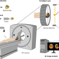
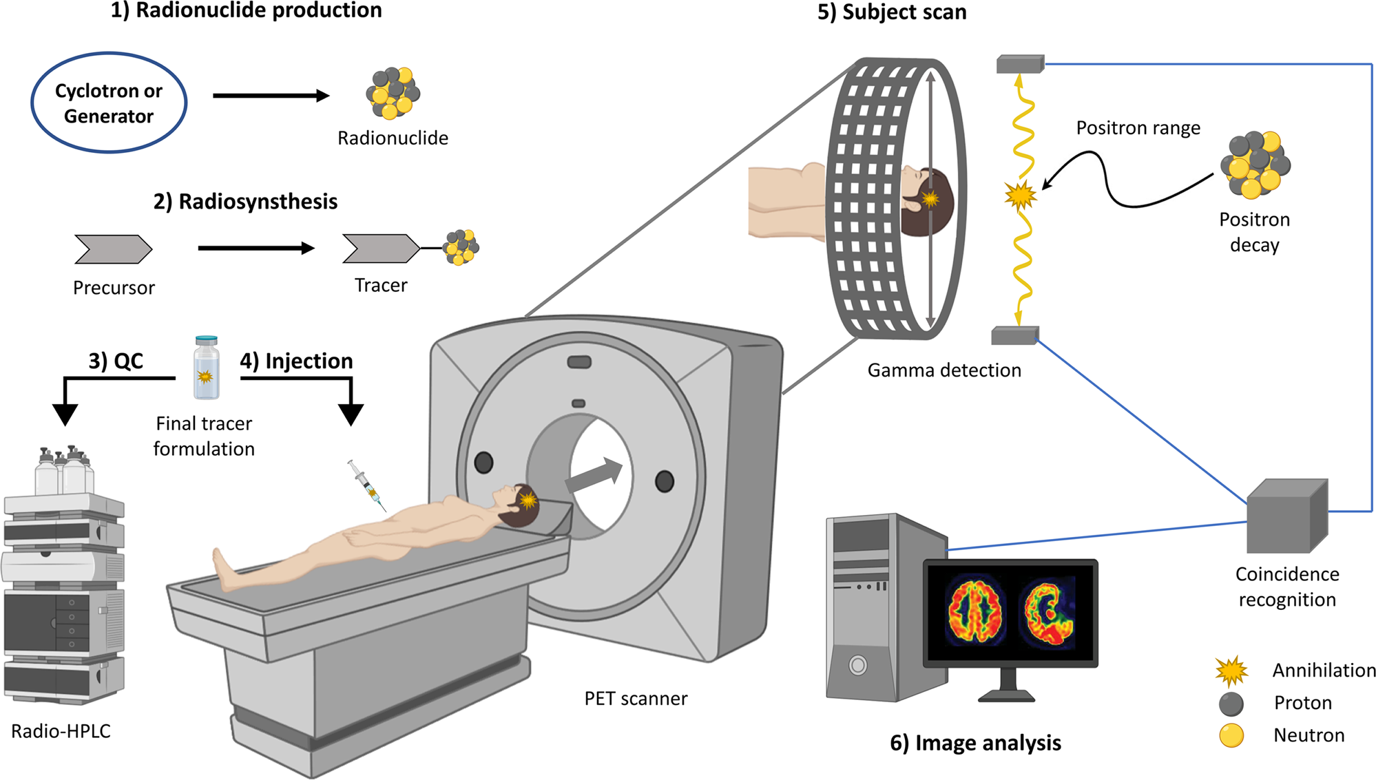
Positron Emission Tomography (PET scan) is an advanced medical imaging technique used to visualize metabolic activity within the body. This technique involves the use of radioactive materials known as “radiotracers,” which are introduced into the body via injection into the bloodstream. The most commonly used radiotracer is Fluorodeoxyglucose (FDG), a radioactive glucose analog used in PET imaging.
PET technology measures the distribution of these radioactive materials in the body to identify areas with increased metabolic activity, which often indicates the presence of biologically active cells such as tumors. After injection, the patient is placed in a PET scanner, which detects the radiation emitted by the radioactive material and converts this information into detailed images showing the metabolic activity within tissues.
How PET Scanning Works to Identify Diseases
Preparation and Injection:
Preparation: Before the scan, patients may be required to fast for a certain period (usually 4-6 hours) to lower blood glucose levels, which helps improve scan accuracy.
Injection: A radiotracer (such as FDG) is injected into a vein. The radiotracer needs some time to distribute throughout the body, typically taking 30-60 minutes to reach optimal tissue distribution.
Imaging:
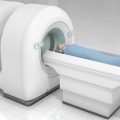
After the waiting period, the patient is placed on a table inside the PET scanner. The scanner passes the patient through and measures the radiation emitted by the radiotracer. This data is collected and converted into three-dimensional images showing the distribution of metabolic activity in the body.
Image Analysis:
After imaging is complete, doctors analyze the resulting images to identify areas with increased metabolic activity, which may indicate the presence of tumors or other conditions. These images provide information about the function of tissues and organs, not just their structure, offering additional insights into the patient’s health.
Importance of PET in Tumor Detection
Early Tumor Detection
The ability to detect tumors early is one of the most significant advantages of PET scanning. The scan can help identify tumors at their earliest stages, even before clinical symptoms appear. Early detection can be crucial for improving treatment outcomes, as it allows for earlier medical intervention and increases the likelihood of successful treatment. Cancerous tissues often have higher metabolic activity compared to healthy tissues, making them take up the radiotracer more and thus appear more prominently in the images.
Assessing Tumor Spread
Once a tumor is diagnosed, determining its spread is vital for developing an appropriate treatment plan. PET scans can provide accurate information about the extent of tumor spread to nearby tissues or other organs. Understanding the extent of the tumor can significantly impact treatment decisions, helping doctors determine whether additional surgery, radiation therapy, or chemotherapy is needed. This information contributes to designing a more effective treatment plan and providing strategies to manage the disease.
Monitoring Treatment Effectiveness
After treatment begins, PET scans can be used to monitor the response of tumors to chemotherapy or radiation therapy. By comparing images taken before and after treatment, doctors can evaluate whether the treatment is improving the condition or if adjustments are needed. This ability to assess treatment effectiveness helps in modifying treatment plans based on the actual response of the tumor, contributing to better treatment outcomes.
Determining Disease Stages
PET scanning is a valuable tool for accurately staging diseases. By providing a precise view of tumor activity, doctors can more accurately determine the stage of the disease. This helps in selecting the most appropriate treatment and providing tailored care that matches each stage of the disease.
Improving Treatment Strategies
With the precise information provided by PET scans, doctors can refine and tailor treatment strategies based on each patient’s condition. For example, if images show increased metabolic activity in specific areas, treatment can be adjusted to target these areas more effectively.
Comparison Between PET and Other Diagnostic Methods
PET vs. X-ray Imaging
PET scanning differs significantly from X-ray imaging. X-ray imaging focuses on creating images of the skeletal structure and organs using X-rays. In contrast, PET scanning focuses on metabolic activity within cells. This means PET can detect diseases at earlier stages, even when structural changes are not visible on X-rays. Therefore, PET is a complementary tool to X-ray imaging, providing additional information about tissue function.
PET vs. Magnetic Resonance Imaging (MRI)
PET scanning also differs from Magnetic Resonance Imaging (MRI) in the type of information it provides. MRI provides detailed images of soft tissues and internal structures, making it useful for anatomical detail. PET scanning, however, focuses on metabolic activity, making it valuable for diagnosing diseases with high metabolic activity, such as tumors. Both scans can be used together to provide a comprehensive view of medical conditions, as each provides different types of information.
PET vs. Computed Tomography (CT)
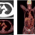
PET scanning and Computed Tomography (CT) complement each other in assessing medical conditions. CT provides detailed images of internal body structures, while PET focuses on metabolic activity. Combining information from both scans can offer a comprehensive view of the medical condition. For instance, CT imaging can locate the tumor, while PET provides information on its metabolic activity, aiding in the development of a cohesive treatment plan.
Summary of What is Positron Emission Tomography (PET) and How It Contributes to Disease Diagnosis
Positron Emission Tomography represents a valuable diagnostic tool that significantly contributes to early disease detection and accurate patient assessment. By providing precise information about cellular metabolic activity, PET helps improve treatment and recovery chances and is essential for evaluating and customizing treatments for each patient.
At Dokki Scan , we offer Positron Emission Tomography services with the highest levels of quality and technology. We are committed to providing the best medical care to our patients using the latest techniques to ensure patient comfort and obtain accurate, reliable results. Our team of specialized doctors and trained technicians is ready to provide support and guidance at every step of the scan, ensuring a smooth and effective experience. For more information about our services, please visit our website: dokki-scan.com.
Latest Blogs
- All Posts
- Blog


