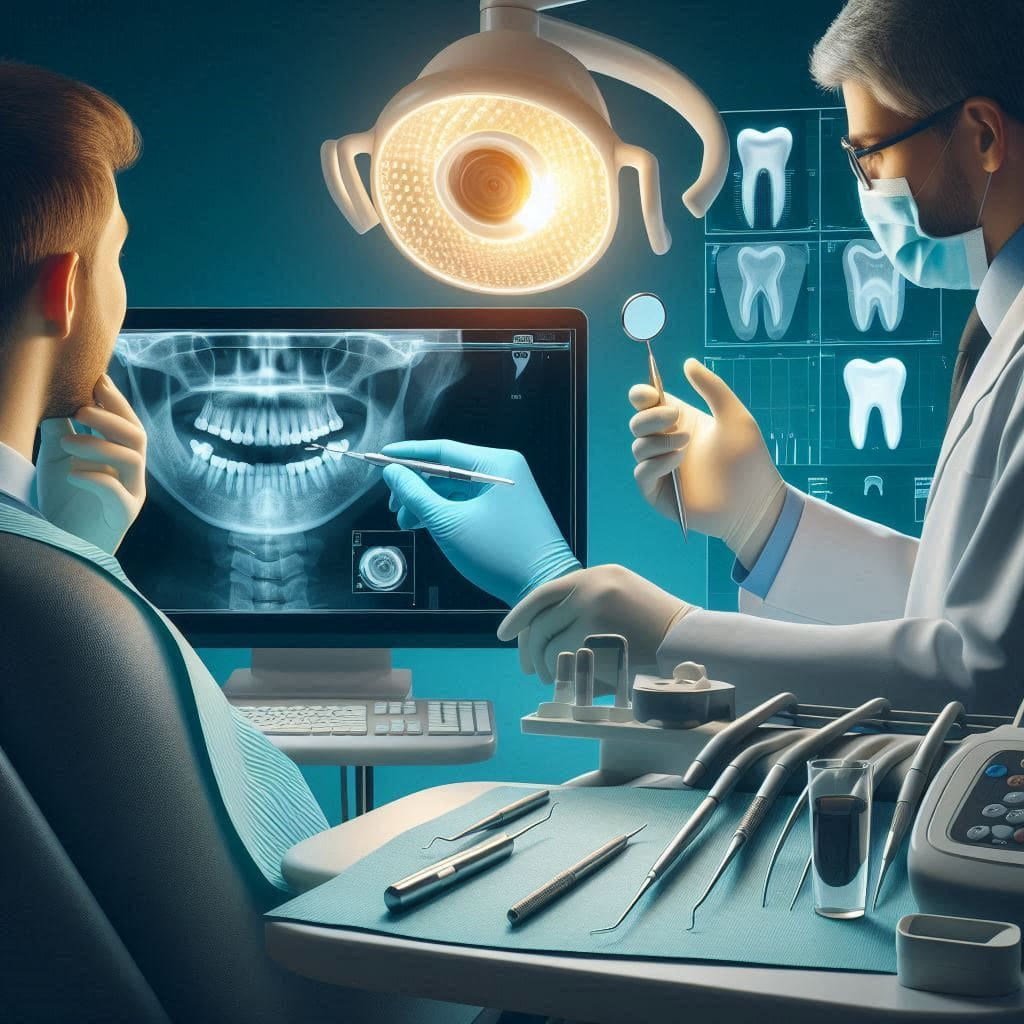
What Are the Types of Dental X-Rays?
What Are the Types of Dental X-Rays? And How Do They Help in Diagnosing Dental Problems?
In the modern world of dentistry, X-rays have become an integral part of the diagnostic and treatment process. This technology allows doctors to see what the naked eye cannot, contributing to better and more accurate patient care. At “Dokki Scan” we offer the latest X-ray technologies to ensure accurate diagnosis and effective monitoring of various cases. In this article, we will explore the different types of dental X-rays and how they are used to diagnose and treat dental issues.
Traditional X-Rays
Definition of X-Rays
X-rays are a type of electromagnetic radiation that can penetrate tissues and capture images of internal body parts. X-rays are widely used in dentistry to detect issues that are not visible to the naked eye.
Types of X-Rays
Panoramic X-Rays
Panoramic X-rays are one of the most commonly used types of X-rays in dentistry. This technique captures a complete image of the mouth, including the teeth, jaws, and surrounding tissues.
Advantages:
Provides a comprehensive image of the mouth.
Helps in detecting impacted teeth, tumors, and jaw abnormalities.
Used in planning surgical treatments such as dental implants.
Uses:
General assessment of dental health.
Detection of major issues that may require surgical intervention.
Assisting in the planning of orthodontic treatments.
Periapical X-Rays
Periapical X-rays focus on a specific area of the mouth, typically one tooth or a small group of teeth.
Advantages:
Provides detailed images of teeth, roots, and surrounding bone.
Helps in detecting cavities, root infections, and bacterial diseases.
Uses:
Diagnosing deep cavities.
Assessing root health.
Assisting in planning root treatments.
Bitewing X-Rays
Bitewing X-rays are used to capture images of the upper and lower teeth in the same area of the mouth.
Advantages:
Provides a view of the teeth from a specific angle.
Helps in detecting cavities between teeth.
Uses:
Identifying interproximal caries (cavities between teeth).
Monitoring the fit of dental restorations.
Benefits of Traditional X-Rays
Accuracy in Detecting Cavities and Root Infections
X-rays are a precise tool for detecting cavities and root infections that may not be visible to the naked eye. Their high accuracy allows doctors to identify the extent of decay and take necessary corrective measures early on.
Role in Identifying Affected Teeth
X-rays assist doctors in pinpointing affected teeth, whether they are fractured, decayed, or experiencing other issues. This enables prompt and effective treatment.
Uses of Dental X-Rays in Disease Diagnosis
Diagnosing Cavities and Gum Diseases
How X-Rays Are Used to Detect Cavities in Their Early Stages
X-rays are effective in detecting cavities in their early stages, identifying decay that has not yet reached the tooth’s surface. This allows doctors to intervene early and address the issue before it worsens.
Role of X-Rays in Assessing the Extent of Gum Diseases
X-rays help evaluate gum health and determine the extent of gum diseases. By visualizing the bone surrounding the teeth, doctors can assess the progression of the disease and take necessary actions.
Diagnosing Tooth and Root Fractures
Using X-Rays to Identify Fractures and Cracks in Teeth and Roots
X-rays are vital for diagnosing fractures and cracks in teeth and roots. Detailed images help doctors determine the location and size of fractures and develop an appropriate treatment plan.
Treatment Planning
Role of X-Rays in Developing Treatment Plans, Such as Orthodontics and Implants
X-rays assist in planning advanced treatments such as orthodontics and dental implants. By providing accurate images of the mouth and teeth, doctors can create a customized treatment plan for each patient.
Monitoring Treatment Progress
How X-Rays Are Used to Monitor Treatment Progress and Ensure Effectiveness
After starting treatment, X-rays are used to monitor progress and ensure effectiveness. Continuous monitoring ensures that treatment is on track and that the patient receives the best possible results.
Modern Digital X-Rays
Definition of Digital X-Rays
Digital X-rays are a modern technology that uses digital devices to capture images instead of traditional film. This technique offers high precision and the ability to process images easily.
Advantages
Reduced Radiation Exposure
One of the main benefits of digital X-rays is reduced radiation exposure for the patient. This makes digital X-rays a safer option for patients, especially children and the elderly.
Image Precision
Digital X-rays provide high-resolution images that can be magnified and analyzed more effectively. This assists doctors in more accurate diagnosis and decision-making.
Quick Results
Thanks to digital technology, X-ray results can be obtained very quickly, speeding up the diagnostic and treatment process.
Uses
Orthodontics
Digital X-rays are widely used in orthodontics to provide accurate images of the jaw and teeth. This helps doctors create detailed treatment plans and achieve better results.
Dental Implants
In dental implants, digital X-rays are used to precisely locate implants and assess the health of surrounding bone and teeth.
Three-Dimensional X-Rays (CBCT)
Definition of Three-Dimensional X-Rays
Three-dimensional X-rays (CBCT) is an advanced technique that provides three-dimensional images of the mouth, jaw, and teeth. This technique offers detailed and comprehensive information that enhances diagnosis and treatment.
Advantages
Providing Three-Dimensional Images
Three-dimensional X-rays provide comprehensive images of the mouth from multiple angles, aiding in more accurate and detailed diagnosis.
Bone and Tissue Analysis
With its ability to analyze bone and tissue in detail, three-dimensional X-rays are used to assess jaw health and plan surgeries more effectively.
Uses
Surgical Planning
Three-dimensional X-rays are widely used in planning complex surgeries, such as dental implants and jaw surgeries. This technique provides detailed images that help doctors develop precise treatment plans.
Evaluating Jaw Health
In orthodontics, three-dimensional X-rays are used to assess jaw health and determine the progress of treatments accurately.
Fluoroscopy
Definition of Fluoroscopy
Fluoroscopy is a technique that uses continuous X-rays to capture live images of internal organs while in motion. This technique is widely used in dentistry to evaluate the dynamics of the mouth and jaw.
Advantages
Providing Live Images
Fluoroscopy provides live images of organs while in motion, aiding in more accurate diagnosis of dynamic issues.
High Precision
Fluoroscopy offers high-resolution images, assisting in accurate diagnosis and effective decision-making.
Uses
Diagnosing Jaw Joint Issues
Fluoroscopy is used to diagnose jaw joint issues and assess its movement accurately.
Planning Orthodontic Treatments
It is also used in orthodontics to evaluate the movement of teeth and jaw accurately and to develop effective treatment plans.
Summary
Dental X-rays, in their various forms, are essential tools in dentistry, playing a significant role in diagnosis and treatment. At “Dokki Scan” we are committed to offering the latest X-ray technologies to ensure the health and safety of our patients. Through X-rays, we can provide accurate diagnosis and effective treatment plans for each case.
Invitation to Visit Dokki Scan
At “Dokki Scan,” we offer a wide range of diagnostic and treatment services using the latest X-ray technologies. If you have dental issues or need a routine check-up, we invite you to visit our website at dokki-scan.com or contact us to schedule an appointment. Our team of experts is ready to provide support and answer all your queries. We are here to ensure your health and safety, and we are committed to providing the best care for you and your family.
Latest Blogs
- All Posts
- Blog